Day 1 :
Keynote Forum
Dionisio Figueiredo Lopes
Hospital de Urgencia Otavio Lage, Brazil
Keynote: The use of multislice CT angiography in the surgical treatment of ruptured intracranial aneurysms
Time : 10:45-11:30

Biography:
Dionisio Figueiredo Lopes is a Neurosurgeon Member of Brazilian Neurosurgery Society, member of Brazilian Neurosurgery Academy. He is the Head of Neurosurgery at Hospital de Urgências Governador Otavio Lage (HUGOL), a hospital reference in neurosurgical emergencies, Consultant at Hospital de Urgências de Goiânia and Hospital Santa Mônica. He is a Neurosurgeon with expertise in vascular diseases, brain tumor and traumatic brain injury. He has Fellowship in Neuro-oncology at Dresden/Germany and Fellowship in Advanced Techniques in Neurosurgery at Tubingen/Germany.
Abstract:
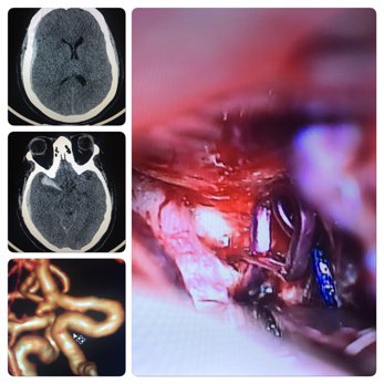
Non-traumatic subarachnoid hemorrhage (SAH) is a neurological emergency. The main cause of non-traumatic SAH (80% of cases) is rupture of an intracranial aneurysm, an event accompanied by high morbidity and mortality rates. The incidence of aneurysmal SAH is estimated to be about 11 cases per 100,000 population per year. Extensive evidence is available demonstrating that early surgery is associated with improved outcome. Cerebral angiography (CA), computed tomography angiography (CTA) or MR angiography are commonly used to determine the location, size and shape of an aneurysm before treatment. CTA images show cerebral vessels in three-dimensional directions and can provide 3D images for aneurysm detection. Some studies have reported sensitivities ranging from 77 and 100% and specificities ranging from 79 and 100%. Among aneurysm detected on CTA and then undergoing surgery, 100% correlation was observed between CTA and CA. CTA, as less invasive and rapidly performed is an accepted method for detection and characterization of cerebral aneurysm when planning surgical intervention. Hospital de Urgências Governador Otávio Lage – HUGOL is a reference hospital for neurological emergencies such as trauma and stroke in a big city in Brazil. We proceeded 60 microsurgical clipping of ruptured intracranial aneurysm during 17 months from August 2015 to December 2016. After the clinical and image diagnoses of SAH, all the 60 patients underwent CTA examinations. The CTA study was performed with a 16-row multislice CT machine. One aneurysm (1.6%) was not detected by CTA initially and visible on the CA. 59 (98.3%) patients were successfully treated based on CTA as the only preoperative investigation. In conclusion, 16-slice CTA image is useful for the diagnosis of ruptured cerebral aneurysm as a noninvasive imaging technique providing an early diagnosis.
Keynote Forum
Antonio Scilimati
University of Bari, Italy
Keynote: Neuroinflammation: Prodrome of neurological and neurodegenerative diseases

Biography:
Antonio Scilimati graduated cum laude in Chemistry at the University of Bari (Italy) and PhD at the University of Wisconsin (USA). He worked for 4 years at MerckSerono plant producing recombinant drugs. Now, he is an Associate Professor at University of Bari, teaching Medicinal Chemistry. In "Medicinal Science", he uses the theranostic approach to target the cyclooxygenase (COX)-1 as a novel biomarker in oncology and neuroinflammation.
Abstract:
Brain inflammatory response, termed neuroinflammation, is crucial to protect the CNS. However, uncontrolled or prolonged neuroinflammation is harmful and could induce neuronal damage. This is particularly relevant in neurological and neurodegenerative diseases (i.e., Alzheimer and Parkinson diseases, amyotrophic lateral sclerosis, multiple sclerosis, traumatic brain injury, HIV dementia, and prion diseases), which are typified by evidence of microglial activation and neuroinflammation. Microglia, the resident immune cells in the brain, plays a role in immune surveillance. Once exposed to immunological challenges such as invading pathogens and neuronal injuries, microglia readily activate and undergo changes in morphology (hypertrophy), number (proliferation), and function (phagocytosis). As a consequence of their activation, microglia produce many pro-inflammatory factors and neurotoxic mediators including complement, arachidonic acid and its lipid metabolites (prostaglandins), cytokines, chemokines, nitric oxide and free radicals, several of which contribute directly to neuronal injury. Among the mechanisms involved into the neuroinflammatory complex network, the cyclooxygenase-1 (COX-1) (predominantly localized in microglia) plays a previously unrecognized role in the neuroinflammation as demonstrated by the attenuation of the inflammatory response and neuronal loss due to the genetic ablation or pharmacological inhibition of COX-1 activity. COX-2, the other known COX isoform, mainly localized in pyramidal neurons, is expected to predominantly contribute to increase prostaglandin biosynthesis in response to insults that directly challenge neurons, such as ischemia and excitotoxicity. In this context, the action of highly selective COX-1 inhibitors compared to coxibs (selective COX-2 inhibitors) in in vitro and in vivo neuroinflammatory state will be presented.

Keynote Forum
Hanan Sheikh Ibrahim
Cleveland Clinic Abu Dhabi, UAE
Keynote: Pseudobulbar affect, cognitive dysfunction and depression in poorly controlled diabetes

Biography:
Hanan Sheikh Ibrahim is a Clinical Assistant Professor at the Cleveland Clinic Lerner College of Medicine of Case Western Reserve University, Ohio, a Consultant Physician and a Quality Officer at the Cleveland Clinic Abu Dhabi. She was trained at Cleveland Clinic in Ohio, USA under the tutelage of Dr. Robert Palmer, Concept Originator of the Acute Care of Elderly (ACE) unit which was modeled internationally. Then she pioneered in the geriatric care in the UAE by establishing the first MACE unit and the first Geriatric Core Curriculum for resident physicians in training. She received her MD from Damascus University, Syria where she specialized in Pulmonary Medicine then she moved to US where she completed her residency in Internal Medicine at the University of Pittsburgh School of Medicine in Pittsburgh, Pennsylvania, US. She completed her Fellowship in Geriatric Medicine at Cleveland Clinic, Ohio. She is board certified in Internal & Geriatrics Medicine.
Abstract:
This study is a case of unrecognized neuro cognitive disorder and pseudo-bulbar affect in a patient with multiple vascular risks, poorly controlled diabetes with subcortical lacunes masquerades as depression treated with Mirtazepin. 64 year old ex-smoker male with PMH of hypertension, long standing poorly controlled type 2 diabetes was presented with insomnia and depression, poorly controlled diabetes where trial of SNRI was partially helpful in his mood but did not help with his crying bursts. He is still driving with multiple episodes of loss of consciousness due to hypoglycemia. His physical exam was unremarkable except for emotional burst of laughter and crying that did not affect congruence. On CGA, he was found to have 3 impaired IADL domains (ability to drive with many car accidents, difficulties handling finances, inability to handle medication, MMSE test - 26/30, low education level, impaired clock drawing test and impaired trail B test, impaired speed, attention and executive skills, GDS was 4/15, FRAIL scale was 4/5. His labs revealed HBA1C above 10. He has normal B12, and folate. His MRI revealed white matter disease, pontine infarct, left thalamic lacunar infarct and left lenticular lacune in addition to cortical atrophy. Patient was recognized as early vascular dementia with associated pseudo bulbar affect masked by depressive symptoms. The case triggered a change of his holistic care that revamped his HbA1C goals and advanced care planning. In summary, general psychiatrists and primary care clinicians may fail to recognize pseudo bulbar palsy and cognitive dysfunction during clinic visits using routine history and physical pseudobulbar affect (PBA), appearing as abrupt episodes of uncontrollable laughter or crying that are incongruent or independent of mood, occurs in many neurological brain diseases or following brain injury. It is important to identify PBA as a different entity from depression, treat and identify underlying vascular cognitive important
- CNS Disorders | Neurosurgery

Chair
Dionisio Figueiredo Lopes
Hospital de Urgencia Otavio Lage, Brazil

Co-Chair
Khin Bo Maung
Northern Lincolnshire and Goole NHS Foundation Trust, United Kingdom
Session Introduction
Debabrata Mukhopadhyay
Kailash Group of Hospitals, India
Title: Challenges in awake craniotomy for intrinsic brain tumors in eloquent cortex
Biography:
Debabrata Mukhopadhyay is currently working as a Neurosurgeon in the Department of Neurosurgery at Kailash Group of Hospitals, India. He has published several articles in the reputed and peer-reviewed journals and participated in several scientific events.
Abstract:
Introduction: Surgical treatment of brain tumors in the eloquent areas has high risk of eloquent impairment. These tumors represent a unique challenge as most of the patients have a higher risk of treatment related complications. Awake craniotomy is a useful surgical approach to help to identify and preserve functional areas in the brain and maximizes tumor removal and minimizes complications.
Methods: Selected patients admitted with intrinsic brain tumor between from July, 2011 to August, 2016 in the eloquent area of brain like speech or motor area were chosen for awake craniotomy. A retrospective analysis was done. A preoperative assessment was also done. These patients were presented with seizure and/or progressive neurological deficit like speech or motor. A standard anaesthesia monitoring was done during surgery. Long acting local anaesthesia (Bupivacaine) was used for scalp block. The surgeries were performed in a state of sleep-awake-sleep pattern, keeping the patients fully awake during tumor removal. Propofol and Fentanyl was used as anaesthetic agents which was completely withdrawn prior to tumor removal. The speech and motor functions were closely monitored clinically by verbal commands during tumor resection. No brain mapping was performed due to lack of resources. All patients underwent non-contrast computed tomogram head in the first post-operative day.
Results: A total of 35 patients were included in the study. The oldest patient was 55 years and youngest being 24 years (mean 36 years). Twenty (57.14%) were females and 15 (42.85%) males. Twenty (57.14%) patients were presented with predominantly seizure disorders and rest with progressive neurological deficit like speech or motor. Thirty (85.71%) patients were discharged on second postoperative day. Complications was encountered in 4 (11.42%) patients who developed brain swelling intraoperatively and 5 (14.28%) deteriorated neurologically in the immediate postoperative period however managed successfully and discharged in a week’s time. Five (14.28%) patients require ICU/HDU care for different reasons. There was no mortality during the hospital stay. Histopathology revealed 25 (71.42%) patients had low grade glioma, 8 (22.85%) had high grade glioma and 2 (5.71%) had metastases.
Conclusion: Awake craniotomy is a safe surgical management for intrinsic brain tumors in the eloquent cortex although surgery and anesthesia is a challenge. It offers great advantage towards disease outcome. However long follow up and more studies are required.
Biography:
Brahim Gargouri is currently working in the Laboratory of Toxicology-Microbiology and Environmental Health at University of Sfax, Tunisia. He has published several original research papers in the reputed and peer reviewed journals.
Abstract:
Substantial evidence has shown that exposure to pyrethroid pesticides may cause adverse neurodevelopmental outcomes and cognitive impairment, but the underlying neurobiological mechanism is poorly understood so far. In this study, we investigated the alterations of neuronal damage, glial activation oxidative stress and cholinergic dysfunction, and their causal relationship with the cognitive deficits induced by bifenthrin. Our results revealed that exposure to bifenthrin for 8 weeks at doses 6 and 21 mg/kgbw leads to reduction in the levels of acetyl-cholinesterase, Na+/K+, Ca2+, Mg2+ ATPases, enzymatic and non-enzymatic antioxidants activities in the hippocampus region. Further, in hippocampus tissue, bifenthrin significantly enhance the mRNA gene expression of nuclear receptor related 1 protein (nurr1), nuclear factor erythroid 2 (nrf2) and nuclear factorkB pathway (NFkB). Oxidative/nitrosative stress was evident in bifenthrin-treated groups by increased malondialdehyde (MDA), protein carbonyls (PCO), and nitrite concentration (NO) in hippocampus. Further, we found that treated rats with bifenthrin exhibited spatial learning and memory impairments and working memory dysfunction compared with control rats. This is also supported by histopathological findings of hippocampus region of rats. Correlational analyses revealed that spatial learning and memory impairments and working memory dysfunction were significantly correlated with the measures of neuronal damage, cholinergic dysfunction and oxidative damage in the hippocampus of treated rats. Moreover, the measures of neuronal damage and central cholinergic dysfunction were significantly correlated with the indexes of oxidative damage in treated rats. The results of the present study suggest that neuronal damage, cholinergic dysfunction and oxidative damage in the hippocampus following bifenthrin exposure could be involved in cognitive deficits.
- Cognitive Neurology | Case Reports

Chair
Antonio Scilimati
University of Bari, Italy

Co-Chair
Hanan Sheikh Ibrahim
Cleveland Clinic Abu Dhabi, UAE
Session Introduction
Melnyk Nataliia O
O O Bogomolets National Medical University, Ukraine
Title: Reactive cuprizone-induced changes in neurons of central nervous system, behavioral reactions and its recovering after influence of leukemia inhibitory factor in mice of different ages

Biography:
Melnyk Nataliia O was investigating demyelination and remyelination process in central nerve system in experiment animals (in rats and mice). She studied structural changes in organs central and peripheral immune system in demyelination condition. She has 262 scientific works (4 patents) and is working in National O O Bogomolets Medical University where she provides lecture courses in Histology, Cytology and Embryology. She has prepared and edited textbook, “Histology, Cytology, Embryology”.
Abstract:
Statement of the Problem: Investigation work was aimed at studying the features of neuroprotective effects of recombinant human leukemia inhibitory factor (rhLIF) on mice of different ages with cuprizone model of demyelination.
Methodology & Theoretical Orientation: In 129/Sv mice at 3-5 and 16-17 months of age, after staining histological sections of brain and spinal cord toluidin blue, were determined the percentage of neurons with unmodified, moderate and severe structural changes. Motor and emotional activity in “open field” test, activity of brain antioxidant enzymes and macrophages capable to phagocytosis of latex beads were assessed. Cuprizone was fed daily for 3 weeks. RhLIF injected after 7 days cuprizone diet, daily, 50 µg/kg. In cuprizone-treated mice of both age groups, increase in the brain and spinal cord proportions neurons with severe changes was observed.
Findings: In young animals, which received cuprizone and rhLIF reduces the amount of neurons with destructive changes. Such changes under influence of rhLIF are slowly observed in older mice. Cuprizone decreases the amount of crossed squares and faecal boluses in mice of both age groups. Inhibition amount and activity of macrophages after injections of the rhLIF presents only for older mice. LIF may be perspective neuroprotective drug in multiple sclerosis. The injections of rhLIF restore emotional activity in these mice, but the increase in motor activity is observed only in young mice. In brain of cuprizone-treated mice of different ages inhibited the activity of catalase and glutathione peroxidase (GP); changes were more pronounced in older mice. The positive effect of rhLIF on GP activity appears only in young mice. Percentage of active macrophages increases in cuprizone-treated mice of both age groups, but their activity is only in 16-17 month-old mice.
Seyed Behnamedin Jameie
Iran University of Medical Sciences, Iran
Title: Neuroprotective effect of exogenous melatonin on the noradrenergic neurons of adult male rats’ locus coeruleus nucleus following REM sleep deprivation
Biography:
Seyed Behnamedin Jameie is currently working as a Professor in the Neuroscience Research Center (NRC) at Iran University of Medical Sciences, Tehran, Iran. He has published several original research papers in the reputed & peer reviewed journals and also participated into several scientific meetings.
Abstract:
Background: Melatonin primarily secrets by the pineal gland in dark phase of the circadian rhythm. In addition to its role as an internal sleep facilitator, melatonin acts as an antioxidant, anti-inflammatory and neuroprotective agents. Recently, melatonin has been introduced as a therapeutic strategy for sleep disorders. Hence, in the present study, we studied the neuroprotective effects of pre- and post-treatment of melatonin in locus coeruleus nucleus (LC) of rapid eye movement (REM) sleep deprived (REM-SD) male adult rats.
Methodology: Adult male rats of control, sham and trial groups were used in this study. By using flower-pot technique, short term REMSD was induced. Exogenous melatonin (ExMe) was used intraperitoneally in two forms of pre and post treatment. The protein level of cleaved caspase-3, number and density of tyrosine hydroxylase (TH) positive neurons and microglia population in LC was studied by Western blot and immunohistochemistry respectively. Morphological changes of LC nucleus and its neurons were also studied by using stereological analysis.
Results: The number of neurons and volume of LC was reserved in animals received post-RSD ExMe, apoptosis significantly was decreased comparing to RSD and Pre-RSD animals. Melatonin post-treatment of RSD rats also decreased cleavage of caspase-3 and increased reduced glutathione content in LC. Moreover, immunohistochemistry analysis revealed the increase number of TH positive neurons and decrease microglia migration.
Conclusion: Based on our findings, antioxidant properties of exogenous melatonin could play a critical role in certain type of sleep disorders.